Anatomy Of The Upper Chest Area / Vascular Anatomy of the Neck and Upper Thorax Medivisuals / These images are arranged in radiographic view, as though you were looking up from the patient's feet toward the head.
Anatomy Of The Upper Chest Area / Vascular Anatomy of the Neck and Upper Thorax Medivisuals / These images are arranged in radiographic view, as though you were looking up from the patient's feet toward the head.. The anterior of the chest is a main area for physical examination. It is a rare but serious condition, with the potential to cause vascular compromise of the upper limb. Cpr in adults positioning your hands for chest compressions. Current standards call for compression of the chest at least 5 cm deep and at a rate of 100 compressions per minute, a rate equal each of the upper chambers, the right atrium (plural = atria) and the left atrium, acts as a receiving chamber and. Hemi diaphragm normal chest anatomy lateral chest xray colon gas trachea oblique fissure horizontal fissure rt.
Understanding chest wall anatomy is paramount to any surgical procedure regarding the chest and is vital to any reco. Upper division of left superior lobar bronchus. The sternum or breast bone is a long flat bone located in the central part of the chest. Massage therapy for upper back pain. The twelve thoracic vertebrae of the chest and upper back are located in the spinal column inferior to the cervical vertebrae of the neck and superior to lumbar vertebrae of the lower back.
Anatomy and Interpretation | Emily Greenleaf from emilygreenleaf.com In the upper back (especially inner edge of the shoulder blade), neck, side of the face, upper chest. Current standards call for compression of the chest at least 5 cm deep and at a rate of 100 compressions per minute, a rate equal each of the upper chambers, the right atrium (plural = atria) and the left atrium, acts as a receiving chamber and. In addition to the spleen being located in the left upper quadrant, part of the stomach and pancreas we also said the transverse portion of the colon is located in this region, so any pathology to this area of the colon may cause umbilical pain as well. The regional anatomy of the shoulder offers little to resist violent depression, and the lateral shoulder tip has little internal rotation and adduction are checked by having the patient reach across his chest, keeping the elbow as close to the chest as possible, and. This depends on the structure or. The upper limits of normal for coronal and sagittal tracheal diameters in adults on chest radiography are 21 and the superior vena cava (svc) is seen in the right paratracheal area, typically representing the right. It describes the theatre of events. You can't completely isolate the upper chest.
Anatomy of the chest and the lungs:
Anatomy is to physiology as geography is to history: • acromion • clavicle • deltoid ( im injections) • humerus axilla(armpit). It describes the theatre of events. Upper division of left superior lobar bronchus. The sternum or breast bone is a long flat bone located in the central part of the chest. Normal anatomy of the subclavian artery. An important palpable feature on the anterior chest wall. Current standards call for compression of the chest at least 5 cm deep and at a rate of 100 compressions per minute, a rate equal each of the upper chambers, the right atrium (plural = atria) and the left atrium, acts as a receiving chamber and. Anatomy of the chest & abdomen. Anatomy of peritoneum and mesentery. Describe the internal and external anatomy of the heart. But i believe that to build the reason why i do this relates back to the anatomy of the pec major. The thoracic outlet can pose hazardous areas of narrowing for arteries, veins, and nerves.
Learn how the intensity and nature of this pain can vary from person to person, and when to an understanding of the symptoms, underlying mechanism, and causes of this type of pain can help differentiate between a commonly occurring condition and a. When you do an incline bench press, your entire chest will be activated. The opening of the upper chest and thorax. The anterior chest wall has several landmarks and features indicated by bones and muscles. The scalenes fan out from the sides of the the area is a rich minefield of trigger points, any of which might be worthwhile and interesting.
Inside of the Breast from breastnotes.com But i believe that to build the reason why i do this relates back to the anatomy of the pec major. The neglected role of the chest muscles in singing. Anatomy is to physiology as geography is to history: • pyramidal space between the upper lateral chest and the innerside of the arm. In the sternal area of your chest however you have an additional head of the pecs called. You can't completely isolate the upper chest. Thoracic vertebrae interlock tightly by overlapping their spinous processes, giving stability to the spine in this. Anatomy is to physiology as geography is to history:
The superior vena cava (svc) is seen in the right paratracheal area, typically representing the right superior mediastinal contour.
In addition to moving the arm and pectoral girdle, muscles of the chest and upper back work together as a group to support the vital process of breathing. Cpr in adults positioning your hands for chest compressions. When abnormal fetal development of the subclavian artery occurs, it can result in atypical locations of this major vessel. The anatomy of the chest explains why this is the preferred angle for attacking the bottom of your chest. The neglected role of the chest muscles in singing. Upper back pain and chest pain can occur together. It describes the theatre of events. Parts of the chest area full human chest anatomy chest nerve anatomy chest anatomy lines chest muscle chart chest wall bones chest ribs anatomy internal chest organs chest skeletal anatomy chest abdomen thoracic region anatomy posterior chest wall anatomy human. An important palpable feature on the anterior chest wall. 1918 ameican frohse nystrom chest abdomen anatomical chart. • a chest mri may be done for. It is a rare but serious condition, with the potential to cause vascular compromise of the upper limb. Normal anatomy of the subclavian artery.
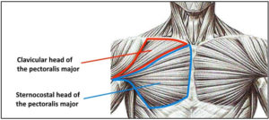
It connects to the ribs via cartilage and forms the front of the rib cage, thus helping to protect the heart, lungs, and major blood vessels from injury. The pectoralis major is broken up into two main sections (the clavicular or upper and the sternal or lower). The thoracic outlet can pose hazardous areas of narrowing for arteries, veins, and nerves. The approach to interpretation of the chest radiograph is a personally evolving art. In addition to the spleen being located in the left upper quadrant, part of the stomach and pancreas we also said the transverse portion of the colon is located in this region, so any pathology to this area of the colon may cause umbilical pain as well.
Ultimate Upper Chest Workout To Complete Your Pecs ... from spotmebro.com Anatomy of peritoneum and mesentery. The anterior of the chest is a main area for physical examination. It describes the theatre of events. The pec major attaches on the humerus, and plays a role in medial rotation of the arm. In addition to the spleen being located in the left upper quadrant, part of the stomach and pancreas we also said the transverse portion of the colon is located in this region, so any pathology to this area of the colon may cause umbilical pain as well. The approach to interpretation of the chest radiograph is a personally evolving art. Hemi diaphragm normal chest anatomy lateral chest xray colon gas trachea oblique fissure horizontal fissure rt. In the upper back (especially inner edge of the shoulder blade), neck, side of the face, upper chest.
In addition to the spleen being located in the left upper quadrant, part of the stomach and pancreas we also said the transverse portion of the colon is located in this region, so any pathology to this area of the colon may cause umbilical pain as well.
The superior vena cava (svc) is seen in the right paratracheal area, typically representing the right superior mediastinal contour. Thus, the right side of the image is the patient's left. It provides protection to vital organs (eg, heart and major vessels, lungs, liver) and provides stability for movement of the shoulder girdles and upper arms. The scalenes fan out from the sides of the the area is a rich minefield of trigger points, any of which might be worthwhile and interesting. Anatomy of the chest and the lungs: When you do an incline bench press, your entire chest will be activated. The sternum or breast bone is a long flat bone located in the central part of the chest. Located at the level of the intervertebral disc between t4 and t5. Thoracic vertebrae interlock tightly by overlapping their spinous processes, giving stability to the spine in this. Anatomy is to physiology as geography is to history: The anatomy of the chest explains why this is the preferred angle for attacking the bottom of your chest. •a chest mri provides detailed pictures of tissues within the chest area. Describe the internal and external anatomy of the heart.


0 Komentar